Jacobs University researchers discover how immune proteins group together
November 23, 2018
The surface of our body cells is covered in proteins that have many tasks. Some of these proteins, the so-called MHC proteins, help the immune system to recognize whether the cell that carries them has become infected by a virus or a bacterium, or whether it has even become part of a tumor. So, the MHC proteins are crucially important to keep us healthy. Researchers at Jacobs University Bremen have now developed a versatile method that can help to understand the interaction of these proteins.
There is one crucial question about MHC proteins that researchers have been unable to answer for the last 25 years: do MHC proteins come together at the cell surface to form groups with specific tasks? MHC proteins, like all surface proteins, are known to float freely at the cell surface like rubber ducks in a pond. They can touch each other, but do these groups play a role in their signaling function? Such questions are very hard to investigate.
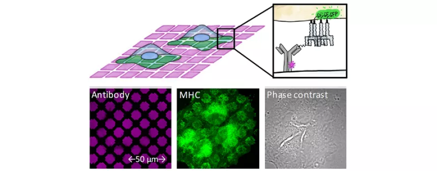
,Now, Dr. Cindy Dirscherl, a biotechnologist and microscopist in the research group of Professor Sebastian Springer at Jacobs University Bremen, has developed a new technique that helps researchers investigate such floating associations of MHC proteins. Cultured cells are seeded on glass plates that have been printed with tiny micropatterns of antibodies, using a special process. The printed antibodies hold on to the MHC proteins and group them into these micropatterns. Some MHC proteins, which have been made fluorescent and introduced into the cell, are then tested for binding to the captured MHC proteins. "If the MHC proteins group together, then we see these green squares in the microscope," says Dirscherl. "We found that only only one form of MHC proteins, the so-called free heavy chain, forms such groups."
The researchers yet do not know what the role of the groups – or clusters, as they call them – actually is, but now that they are identified, they can be investigated in great detail. "One really big advantage of our new method is that it can tell us which exact form of the protein we are looking at. This has never been possible before," says Springer, who offers a view into the future. "The technique that Cindy Dirscherl has developed can be applied to any surface protein. There are so many more exciting and important ones, and we cannot wait to see what people will discover when they use our new technology."
The work was published in the journal eLife:
Cindy Dirscherl, Zeynep Hein, Venkat Raman Ramnarayan, Catherine Jacob-Dolan, and Sebastian Springer:
A two-hybrid antibody micropattern assay reveals cell surface clustering of MHC I heavy chains.
eLife 7 (2018): e34150. doi: 10.7554/eLife.34150
The experimental system explained in a YouTube video:
Questions will be answered by:
Dr. Sebastian Springer | Professor of Biochemistry and Cell Biology
s.springer [at] jacobs-university.de | Tel: +49 421 200-3243
